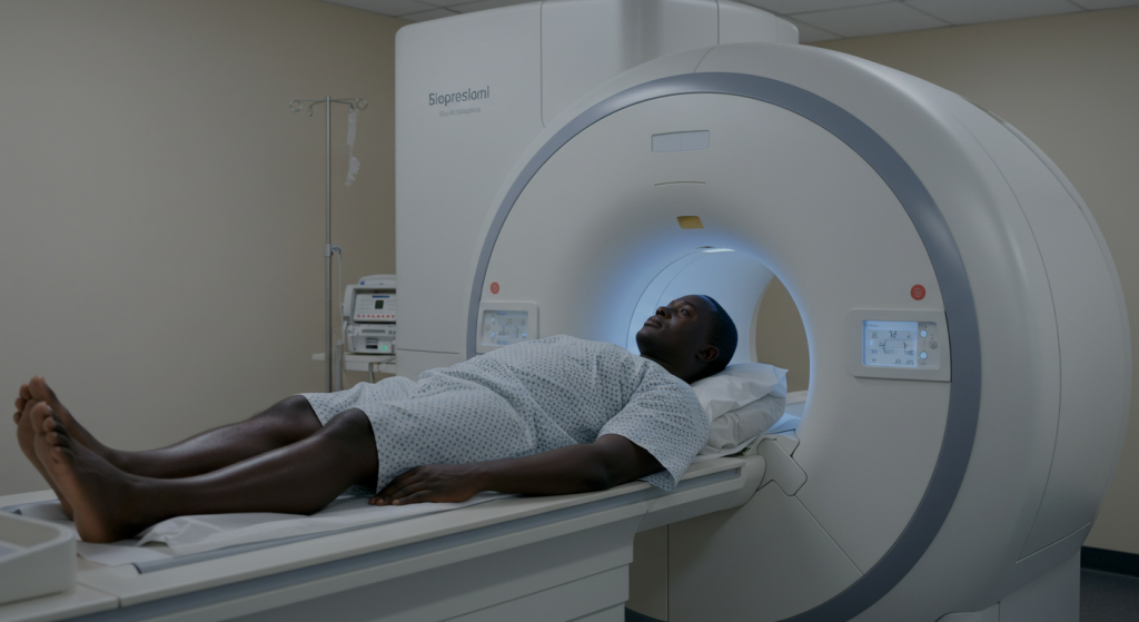
An MRI scan is a marvel of medicine, painting detailed pictures of your insides without a single cut. But for some, sliding into that narrow, tunnel-like machine stirs up a beast called claustrophobia—fear of tight spaces. If the thought of being boxed in makes your heart race, you’re not alone. Plenty of folks dread the MRI not for the results, but for the feeling of being trapped. Understanding what’s behind it can turn a scary moment into something you can handle.
So, what happens? You lie on a bed that glides into a tube—sometimes just wide enough for your shoulders. The machine clanks and hums, and you’re stuck there for 20 minutes or more. For someone with claustrophobia, that closeness can trigger panic—sweaty palms, a pounding chest, even a urge to bolt. It’s not just the space; the noise and stillness pile on. Some machines are older, tighter, or louder, while newer “open” MRIs feel less confining—though they’re not everywhere yet.
The good news? You’ve got options to cope. Tell your doctor ahead of time if tight spots freak you out—they might offer a mild sedative to take the edge off. Bringing a friend to chat with you through the intercom can help too. Technicians often let you pick music or give you a panic button to press if it gets too much. Picture yourself somewhere open—like a beach or a field—while you’re in there. Even a simple eye mask can trick your brain into feeling less caged.
Prep makes a difference. Ask about the machine beforehand—how tight is it, how long will it take? Some places let you peek at it first, easing the shock. Costs vary, and in busy clinics, staff might not coddle you, so speak up about your fears. Afterward, you might feel rattled, but it fades fast—results come soon, and you’re out. It’s not a breeze, but it’s doable with a little planning.
Claustrophobia in an MRI isn’t rare, and it’s not a weakness—it’s just your mind sounding an alarm. Whether it’s your knee, brain, or back being scanned, knowing what sets it off and how to fight back keeps you in charge. Don’t let fear stop you from a test that could save your life—it’s a short tunnel to cross, and you’re tougher than you think.