Magnetic Resonance Imaging (MRI) is a powerful diagnostic tool that uses strong magnetic fields and radio waves to create detailed images of organs and tissues. Unlike X-rays or CT scans, MRI does not use ionizing radiation, making it a safer option for imaging soft tissues such as the brain, muscles, and joints. However, the experience can be intimidating for first-time patients due to the enclosed nature of the machine and the loud noises it produces. Understanding how an MRI works and what to expect can ease anxiety and ensure a smooth procedure.
Before undergoing an MRI, patients must remove any metal objects, including jewelry, watches, and even certain clothing items with metal zippers or buttons. Individuals with implanted medical devices, such as pacemakers or metal prosthetics, should inform their radiologist beforehand, as some materials may interfere with the scan. In some cases, contrast dye is used to enhance image clarity, requiring patients to disclose any allergies or kidney-related conditions to their healthcare provider.
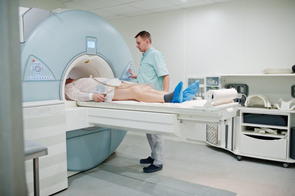
During the scan, patients lie on a motorized table that slides into the MRI machine. The procedure can last anywhere from 15 minutes to over an hour, depending on the area being examined. While the machine itself is not painful, remaining still for extended periods can be challenging for some patients. To reduce discomfort, many imaging centers offer earplugs or headphones with music to help mask the loud tapping and thumping noises produced by the MRI scanner.
Patients with claustrophobia may find the experience particularly difficult, but open MRI machines and mild sedatives are available options to improve comfort. Deep breathing exercises and visualization techniques can also help alleviate stress. Speaking with the technician through the built-in intercom system provides reassurance, and in many cases, patients are allowed to have a companion in the room for support.
Once the scan is complete, there is no recovery time needed, and normal activities can resume immediately unless sedation was used. The radiologist will analyze the images and provide a report to the referring doctor, who will discuss the findings with the patient. By preparing properly and understanding the procedure, patients can undergo an MRI with confidence and ease.