Congenital heart diseases (CHDs) refer to structural abnormalities in the heart that occur during fetal development. These defects can range from mild conditions, such as small holes between heart chambers, to severe malformations requiring immediate medical intervention. Some of the most common CHDs include atrial septal defects (ASD), ventricular septal defects (VSD), tetralogy of Fallot, coarctation of the aorta, and transposition of the great arteries. The severity of CHDs varies significantly, with some defects causing minimal symptoms while others lead to life-threatening complications soon after birth. Symptoms may include abnormal heart murmurs, difficulty breathing, poor weight gain, cyanosis (bluish skin discoloration), and extreme fatigue. Early detection is crucial for optimal management and improved patient outcomes.
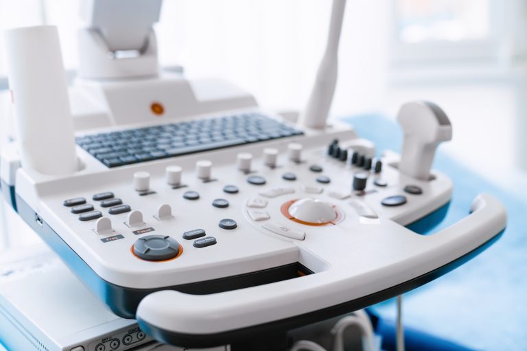
The primary imaging modality for diagnosing CHDs before birth is fetal echocardiography, which uses ultrasound waves to generate detailed images of the fetal heart. This test is typically performed between 18 and 24 weeks of gestation for high-risk pregnancies, such as those with a family history of CHDs or maternal conditions like diabetes. After birth, transthoracic echocardiography (TTE) is the standard imaging method for infants and children. TTE provides real-time visualization of heart structures, blood flow, and valve function, making it an essential tool for confirming CHD diagnoses and assessing severity. Transesophageal echocardiography (TEE), which involves inserting a probe into the esophagus, offers clearer images of deeper heart structures and is often used when transthoracic imaging is inconclusive.
For more detailed anatomical assessment, cardiac MRI (CMR) is increasingly used to visualize congenital defects, particularly in older children and adults with repaired CHDs. CMR provides high-resolution 3D images of the heart and blood vessels without radiation exposure, making it a preferred option for long-term follow-up. CT angiography (CTA), a non-invasive imaging technique involving contrast-enhanced X-ray imaging, is particularly useful for assessing complex heart structures and vascular anomalies. It provides rapid, high-resolution imaging, especially beneficial for pre-surgical planning. In severe cases, cardiac catheterization (angiography) may be necessary, where a thin tube is inserted into the blood vessels to directly measure heart pressures and oxygen levels while obtaining dynamic images of blood flow. This procedure is often used to guide interventional treatments, such as closing septal defects or expanding narrowed arteries.
Advancements in imaging technology have significantly improved the early detection and management of congenital heart diseases. Early diagnosis allows for prompt intervention, which can be life-saving in severe cases. Many congenital heart defects that once required open-heart surgery can now be treated using minimally invasive catheter-based techniques, leading to shorter recovery times and reduced surgical risks. Additionally, continuous monitoring through non-invasive imaging ensures that children and adults with CHDs receive timely treatment and avoid complications such as heart failure or arrhythmias. With ongoing research and technological innovations, the prognosis for individuals with congenital heart diseases continues to improve, offering better quality of life and longer survival rates.
Parents of children diagnosed with CHDs should work closely with pediatric cardiologists and imaging specialists to develop a comprehensive care plan. Regular follow-up imaging and medical supervision are essential to ensure optimal heart function and overall well-being. As medical advancements continue to evolve, the future holds promising improvements in the diagnosis and treatment of congenital heart diseases, offering hope for better patient outcomes and enhanced quality of life.