Doppler ultrasound is a specialized type of ultrasound that measures blood flow in arteries and veins. Unlike standard ultrasound, which creates structural images, Doppler ultrasound detects the movement of red blood cells to assess circulation. It is commonly used to diagnose conditions such as deep vein thrombosis (DVT), peripheral artery disease (PAD), and carotid artery stenosis.
One of the biggest advantages of Doppler ultrasound is that it is non-invasive and does not require contrast dye, unlike traditional angiography. This makes it a safer alternative for patients with kidney disease or those allergic to contrast agents. It is often used to monitor patients at risk for stroke or blood clots, as it can detect narrowing or blockages in the arteries.
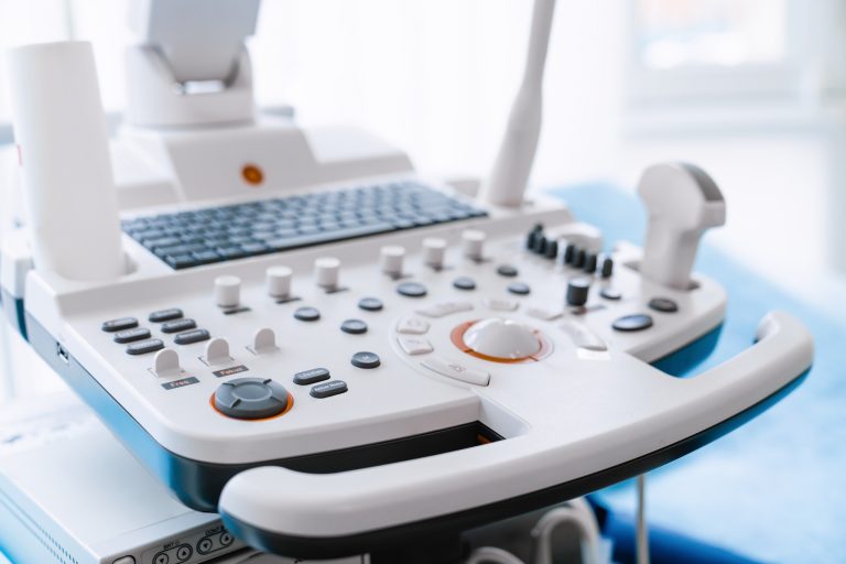
The procedure is similar to a regular ultrasound but focuses on blood flow dynamics. The sonographer applies gel to the skin and moves the transducer over the target area, emitting sound waves that bounce off moving blood cells. The machine translates these reflections into color-coded images, showing the speed and direction of blood flow.
Doppler ultrasound is commonly used to assess varicose veins, leg swelling, and poor circulation. It helps vascular surgeons determine whether a patient needs medication, lifestyle changes, or surgical intervention. For pregnant women, Doppler ultrasound can also evaluate blood flow in the umbilical cord, ensuring the baby receives adequate oxygen and nutrients.
As a quick, painless, and highly informative test, Doppler ultrasound plays a crucial role in diagnosing vascular conditions early. Patients experiencing leg pain, swelling, or dizziness should discuss with their doctor whether a Doppler scan could help assess their circulation health.