Ischemic cardiomyopathy (ICM) is a severe form of heart disease caused by prolonged reduced blood supply to the heart muscle, usually due to coronary artery disease (CAD). Over time, repeated episodes of ischemia (low oxygen supply) weaken the heart, leading to heart failure. Symptoms include shortness of breath, fatigue, chest pain, and swelling in the legs. If untreated, ICM can progress to life-threatening complications like arrhythmias and heart failure.
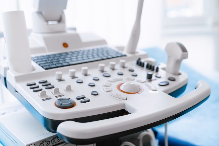
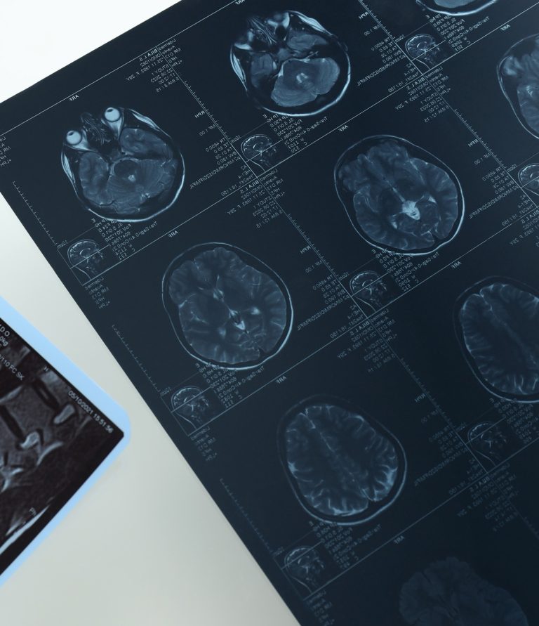
Diagnosis of ischemic cardiomyopathy relies on several imaging techniques. Echocardiography (ultrasound of the heart) is the first-line test, providing real-time images of heart muscle movement and function. It helps assess left ventricular ejection fraction, a key indicator of heart failure severity. Cardiac MRI offers more detailed visualization of heart structure and scarring from previous heart attacks. Coronary angiography (using X-ray and contrast dye) is crucial in determining blockages in coronary arteries and guiding potential interventions like stent placement.
Other imaging tools include CT coronary angiography, which provides non-invasive visualization of coronary arteries, and nuclear stress tests, which evaluate blood flow to the heart under exertion. These imaging techniques help cardiologists determine whether revascularization procedures, such as bypass surgery or stenting, are necessary.
Early detection and intervention can significantly improve outcomes in ischemic cardiomyopathy. Patients with CAD should undergo regular screenings, lifestyle modifications, and medication management to prevent the progression of heart failure.


