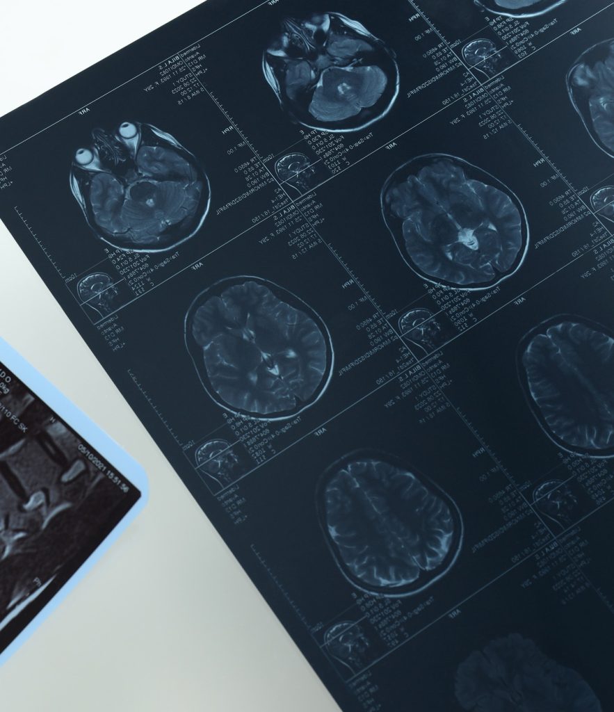
Magnetic Resonance Imaging (MRI) has revolutionized the diagnosis of neurological disorders, providing unparalleled detail of the brain and spinal cord. Unlike CT scans or X-rays, MRI uses strong magnetic fields and radio waves to generate high-resolution images of soft tissues, making it particularly effective for identifying brain abnormalities. Neurologists rely on MRI scans to detect conditions like multiple sclerosis, brain tumors, strokes, and epilepsy, often before symptoms become severe.
One of the most critical applications of MRI in neurology is in detecting multiple sclerosis (MS). MS affects the nervous system by damaging myelin, the protective covering of nerves. MRI scans can reveal these lesions, even in early stages, allowing for prompt treatment that can slow the disease’s progression. Without MRI, MS diagnosis would be much more difficult, as its symptoms often mimic other conditions.
MRI also plays a vital role in identifying strokes. When a patient shows signs of a stroke, doctors must quickly determine whether it is caused by a blood clot (ischemic stroke) or bleeding in the brain (hemorrhagic stroke). MRI provides precise imaging of brain tissue and blood vessels, helping guide emergency treatment decisions that can prevent long-term disability.
For patients experiencing seizures, MRI is often used to detect structural brain abnormalities, such as tumors or malformed blood vessels, which might be triggering the condition. It also helps pinpoint the exact location of abnormalities, assisting neurosurgeons in planning surgical interventions when necessary. In cases of brain tumors, MRI not only identifies the presence of a mass but also provides crucial details about its size, location, and possible impact on surrounding brain structures.
For patients with neurological concerns, MRI provides critical insights without the risks of radiation exposure. While the procedure requires lying still in an enclosed machine, its benefits far outweigh any discomfort, allowing for early detection and better treatment outcomes.