Table of Contents

Need Clarity? Get Personal Guidance.

Not sure what your scan means or how to prepare? Book a one-on-one consultation with our radiology expert.

Ultrasonography Scan

Curious about your upcoming radiology exam? This post provides clear, patient-friendly information to help you understand what to expect, how to prepare, and what your results might mean.
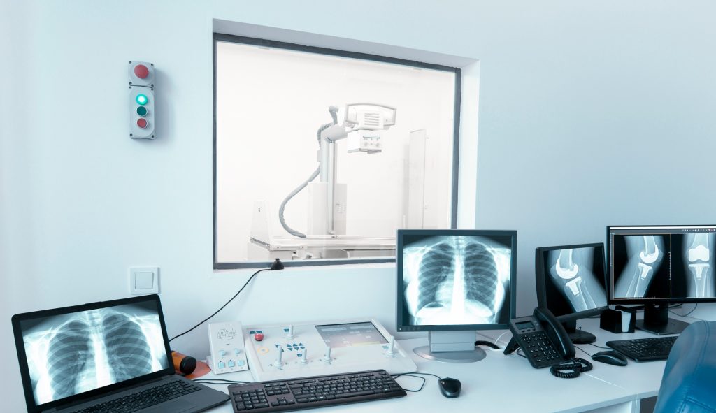
X-ray laboratory with monitors and modern equipment

Introduction

Ultrasound scan, also known as ultrasonography, “scan” (in layman terms) or a sonogram, is a medical imaging technique that uses high-frequency sound waves to produce images of the inside of the body. The sound waves bounce off different tissues, giving off echoes that are captured and turned into real-time images or videos that are viewed on a monitor screen.

Unlike x-rays, ultrasound does not use ionizing radiation to create images. This makes it a safe and non-invasive method for visualizing internal organs, tissues, and even unborn babies. Ultrasound is widely used in Radiology for routine checks, to evaluate conditions such as pain, swelling, or infections and even in emergency situations due to its cost effectiveness and easy accessibility. 

Frequently Asked Questions

What are the common Uses of Ultrasound?

Ultrasound can be used to diagnose a variety of medical conditions and monitor treatment. Some of its most common applications include:

  • Monitoring fetal development during pregnancy.
  • Diagnosing organ conditions like liver disease, gallstones, or kidney stones.
  • Guiding procedures such as biopsies.
  • Evaluating blood flow using Doppler ultrasound to detect blockages or blood clots.

Given its wide range of applications, relative affordability when compared with some other modalities, and safety and convenience, ultrasound remains an invaluable tool in modern medicine.

What are the Types of Ultrasounds we have?

Before diving in here, let us start by explaining how the ultrasound machine has a small handheld device of different shapes called a probe or transducer. This probe will be the one to send in sound waves into the body and also receive returning echoes from your body. 

Depending on how the probe is applied, there are several types of ultrasounds, and oftentimes, the choice depends on your Radiologist, Sonographer or Sonologist, the area being examined and the purpose of the test. The most common types include:

  • External Ultrasound Scan

Here, the ultrasound probe is placed on the skin, over the area to be examined with a gel applied on the skin to allow for better contact, movement across the area, and easy ultrasound signal conduction. This method is used for various organs like the heart, liver, kidneys, and in pregnancy for monitoring the fetus.

  • Internal Ultrasound Scan

In this case, the probe is inserted into a part of the body, such as the rectum (transrectal ultrasound) or vagina (transvaginal ultrasound). This method is used to obtain clearer images of internal organs, such as the prostate (in males) or the uterus and ovaries (in females).

  • Endoscopic Ultrasound: 

This is similar to an internal ultrasound except that in this case, a specialized probe attached to an endoscope is guided into deeper parts of the body, such as the oesophagus, allowing detailed images of these internal areas.

  • Doppler Ultrasound: 

Unlike the other types of ultrasounds which we have mentioned that focus on structures in your body, this type of ultrasound focuses on evaluating blood flow within the body. The knowledge of blood flow can be used to tell what kind of disease process is occurring. 

How do I prepare for an Ultrasound?

Preparation for an ultrasound largely depends on the type of scan being performed: On a general note, 

Before the Scan: 

  • For abdominal scans, such as those for the liver or gallbladder, you may be asked to avoid eating or drinking for several hours to get clear images.
  • For pelvic or prostate scans, you might need to drink water and avoid urinating so that your bladder is full during the exam, making the images clearer.

In some cases, no specific preparation is needed, especially for scans of the heart, thyroid, or soft tissues.

During the Scan: 

  • You will be positioned on an examination bed, and a small amount of gel will be applied to the skin in the area being examined. This gel helps transmit the sound waves into the body.
  • The healthcare professional (Radiologist, sonographer, or sonologist) will move the ultrasound probe over the area to acquire the images of your area of concern.
  • In internal ultrasounds, the probe is gently inserted into the body to provide clearer images of certain internal structures.

After My Scan?

Once your scan is complete, the gel will be wiped off, and you can resume your normal activities. Most ultrasound exams do not require recovery time, and the procedure itself is painless. However, for internal ultrasounds, there may be slight discomfort during the insertion of the probe.

What Are the Risks of Ultrasound?

Ultrasound is considered one of the safest imaging modalities available today because it does not use radiation, unlike x-rays or CT scans. It is also non-invasive and can be routinely used to monitor pregnancies and diagnose various medical conditions.

Can my Child/Newborn Safely have an Ultrasound?

Yes. Ultrasound is safe for both adults and children, including pregnant women, due to the lack of ionizing radiation. There are no known harmful effects associated with standard diagnostic ultrasound.

What form of discomfort will I experience?

While most ultrasound scans are painless, internal ultrasounds can cause minor discomfort during the procedure due to the insertion of the probe. However, this discomfort is usually minimal and short-lived. Always maintain communication with the healthcare professionals carrying out your test, and let them know if you feel very uncomfortable.

Share this article

Continue Reading

No posts found