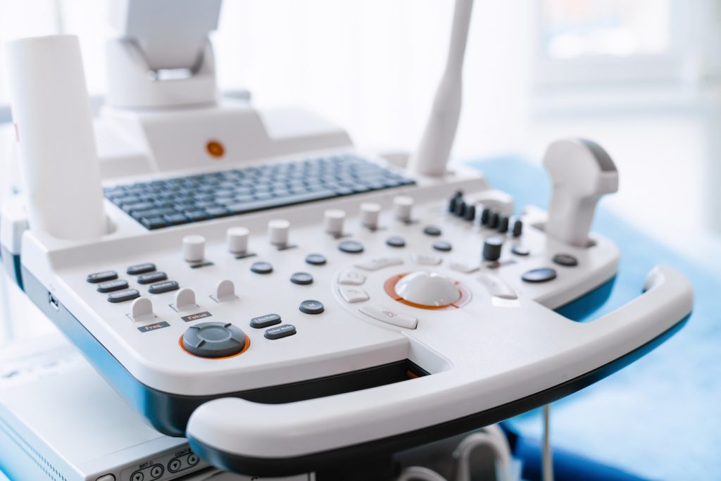
Ultrasound, also known as sonography, is a widely used imaging technique that employs high-frequency sound waves to create images of internal structures. Unlike X-rays and CT scans, ultrasound does not use ionizing radiation, making it a safer option for pregnant women and children. It is commonly used to assess organs like the liver, kidneys, and heart, as well as to monitor fetal development.
One of the greatest advantages of ultrasound is its real-time imaging capability. This allows doctors to observe moving structures, such as blood flow in arteries and veins, heart valve motion, and fetal activity. It is especially useful in emergency settings where quick assessment of internal bleeding or organ damage is required.
Compared to MRI and CT scans, ultrasound is more accessible and cost-effective. Many hospitals and clinics have ultrasound machines readily available, reducing wait times and making it an efficient first-line diagnostic tool. However, ultrasound has limitations—it does not penetrate bone well and is less effective for imaging air-filled organs like the lungs.
A key factor in ultrasound’s effectiveness is the skill of the sonographer. Unlike MRI and CT, which produce standardized images, ultrasound requires an experienced technician to capture and interpret high-quality images. Variability in results can occur depending on patient anatomy and positioning.
Despite its limitations, ultrasound remains one of the safest and most versatile imaging tools available. Whether for pregnancy monitoring, organ evaluation, or guiding minimally invasive procedures, it provides critical information without radiation exposure, making it an invaluable part of modern medicine.


