
RadFAQS
Scan GuidesFind a CenterTalk to a Radiographer
Find/Book
HomeScan GuidesFind a CenterTalk to a RadiographerBooking Follow-Up
RadFAQS

Nigeria's radiology directory - helping patients find listed diagnostic centers, understand their scans, and take control of their health.

Modalities

  • CT Scan
  • MRI
  • DEXA
  • X-ray
  • Ultrasound
  • Fluoroscopy
  • Mammography
  • Nuclear Medicine
  • Interventional Radiology
  • Radiotherapy

Platform

  • Find/Book a Scan
  • Talk to a Radiographer
  • Booking Follow-Up
  • Dictionary
  • Volunteer

Portals

  • Center Login

Stay in the Know

Get updates on new centers, health tips, and platform news.

support@RadFAQS.comWhatsApp Us
© 2026 RadFAQS. All rights reserved.
Privacy PolicyTerms of UseRefund PolicyAbout
Back to Articles
Scan Basics

Pregnancy and Medical Imaging: When Radiation Is a Concern

Written by Oluwatobiloba Akinnusi, B.Sc Radiography
·
Medically reviewed by Olusegun Samuel Faith, M.Sc (Medical Imaging), MPH, PgDip (MRI)· Last reviewed 29 Jul 2026
Pregnancy and Medical Imaging: When Radiation Is a Concern

In this article

Which scans use ionising radiation?The body part makes a differenceWhy pregnancy may change or delay an examinationContrast and nuclear medicine need separate decisionsWhat if you had a scan before knowing you were pregnant?When should you speak up?

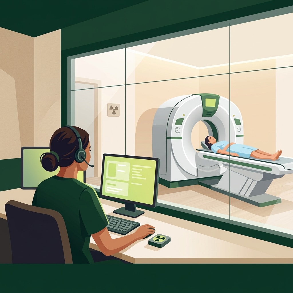
Radiographers often ask whether you are pregnant or could be pregnant before an X-ray, CT, fluoroscopy, or nuclear medicine examination. The question is a safety check, not a declaration that the scan cannot be done.

Pregnancy changes how the team weighs the benefit of the examination, the possible exposure to the developing baby, and whether another type of imaging could answer the same question.

Which scans use ionising radiation?

X-ray, CT, fluoroscopy, and nuclear medicine use ionising radiation. The amount and route of exposure differ between them.

Ultrasound does not use ionising radiation. MRI uses a magnetic field and radio waves rather than X-rays. When either can answer the clinical question, it may be preferred during pregnancy. This does not mean ultrasound or MRI can replace every urgent or necessary X-ray or CT.

The body part makes a difference

An examination of the abdomen or pelvis can expose the pregnancy directly to the X-ray beam. An X-ray or CT of the head, chest, arm, or leg does not place the pregnancy in the direct beam, although a very small amount of scattered radiation may reach it.

The dose also depends on the examination and technique. A single chest X-ray, an abdomen and pelvis CT, and a long fluoroscopy procedure should not be described as though they carry the same exposure.

Why pregnancy may change or delay an examination

If an examination is routine and can safely wait, the referring clinician and radiology team may postpone it or choose ultrasound or MRI. If the result is needed to diagnose a serious illness or injury, delaying the scan may create more risk for both mother and baby.

The team may adjust the scan range or technique, remove unnecessary image phases, or use another examination where appropriate. The decision should be based on the medical question, not fear alone.

Contrast and nuclear medicine need separate decisions

Iodine-based CT contrast and gadolinium-based MRI contrast are not used routinely in pregnancy without considering why they are needed. Gadolinium is generally avoided unless the expected benefit justifies its use.

Nuclear medicine tracers behave differently from external X-rays because radioactive material is introduced into the body. The tracer, dose, timing, and clinical need all matter. This requires case-specific advice from the nuclear medicine team.

What if you had a scan before knowing you were pregnant?

Do not assume harm has occurred. Tell your obstetric clinician and the imaging centre what examination you had and when. If the uterus was exposed, a radiologist or medical physicist can estimate the dose when that information would help.

Ending a wanted pregnancy is not usually recommended solely because a diagnostic imaging examination occurred. The actual exposure and clinical circumstances need proper assessment.

When should you speak up?

Tell the imaging team before the examination if:

  • you know you are pregnant;
  • your period is late or pregnancy is possible;
  • you are receiving fertility treatment or could be in a very early pregnancy; or
  • you are breastfeeding and a nuclear medicine examination is planned.

The centre may ask about the first day of your last menstrual period or recommend a pregnancy test. Local policies vary, especially for examinations involving the abdomen or pelvis.

The bottom line

Pregnancy is not an automatic contraindication to all medical imaging. It is a reason to choose the examination carefully, minimise avoidable exposure, and make sure a necessary diagnosis is not delayed.

Radiology education only

RadFAQS explains radiology terms, scan preparation, and what patients commonly experience. It is not a diagnosis, treatment plan, or replacement for your referring doctor, radiologist, or care team. RadFAQS does not monitor this site for emergencies and cannot respond in real time. If symptoms are severe, worsening, or urgent, do not wait for a reply here — contact a healthcare professional or emergency service immediately.

Related FAQ guides

fluoroscopy

Fluoroscopy and Contrast Media Administration

Introduction to Fluoroscopy Contrast Dyes in fluoroscopy, a special dye called contrast media is sometimes used to help create a clearer view of internal structures like blood vessels, organs, the gastrointestinal tract, or joints. This...

mri

Liver MRI with Hepatobiliary Contrast

A Liver MRI with hepatobiliary contrast is a detailed scan that uses a special dye taken up by liver cells. It can provide highly detailed test for characterising liver lesions and is often used to confirm or rule out liver cancer without a biopsy.

mri

MRI with Contrast (Gadolinium): What to Expect

Many MRI scans need a special dye called gadolinium to highlight tissues and blood vessels. This guide explains what gadolinium is, who should not receive it, what side effects to expect, and why your doctor might ask for a kidney test first.

nuclear-medicine

Nuclear Medicine: General FAQs

Nuclear medicine uses tiny amounts of radioactive tracer and special cameras to show how organs and tissues are working. It helps doctors answer questions that ordinary X-rays, ultrasound, CT, or MRI may not answer on their own.

Related dictionary terms

Term

Contrast

Contrast is a substance used during some scans to help blood vessels, organs, inflammation, or tumors show up more clearly.

Procedure

Fluoroscopy

Fluoroscopy is a real-time X-ray technique that shows motion instead of only a still image.

Procedure

MRI

MRI uses magnets and radio waves to create detailed images, especially of the brain, spine, joints, and soft tissues.

Procedure

Nuclear Medicine

Nuclear medicine uses small amounts of radioactive tracer to show how tissues and organs are functioning, not just how they look.