
RadFAQS
Scan GuidesFind a CenterTalk to a Radiographer
Find/Book
HomeScan GuidesFind a CenterTalk to a RadiographerBooking Follow-Up
RadFAQS

Nigeria's radiology directory - helping patients find listed diagnostic centers, understand their scans, and take control of their health.

Modalities

  • CT Scan
  • MRI
  • DEXA
  • X-ray
  • Ultrasound
  • Fluoroscopy
  • Mammography
  • Nuclear Medicine
  • Interventional Radiology
  • Radiotherapy

Platform

  • Find/Book a Scan
  • Talk to a Radiographer
  • Booking Follow-Up
  • Dictionary
  • Volunteer

Stay in the Know

Get updates on new centers, health tips, and platform news.

Occasional scan guidance and RadFAQS updates. Unsubscribe anytime.

support@RadFAQS.comWhatsApp Us
© 2026 RadFAQS. All rights reserved.
Privacy PolicyTerms of UseRefund PolicyAbout
Back to Articles
Before Your Scan

What Multiple CT Scans Mean for Your Radiation Exposure

Written by Taiwo Oluwayemisi, B.Sc Radiography
·
Medically reviewed by Olusegun Samuel Faith, M.Sc (Medical Imaging), MPH, PgDip (MRI)· Last reviewed 10 Jun 2026
What Multiple CT Scans Mean for Your Radiation Exposure

In this article

CT dose is not one fixed numberWhy doctors still request repeat CTHow to avoid repetition that does not helpChildren need extra attention

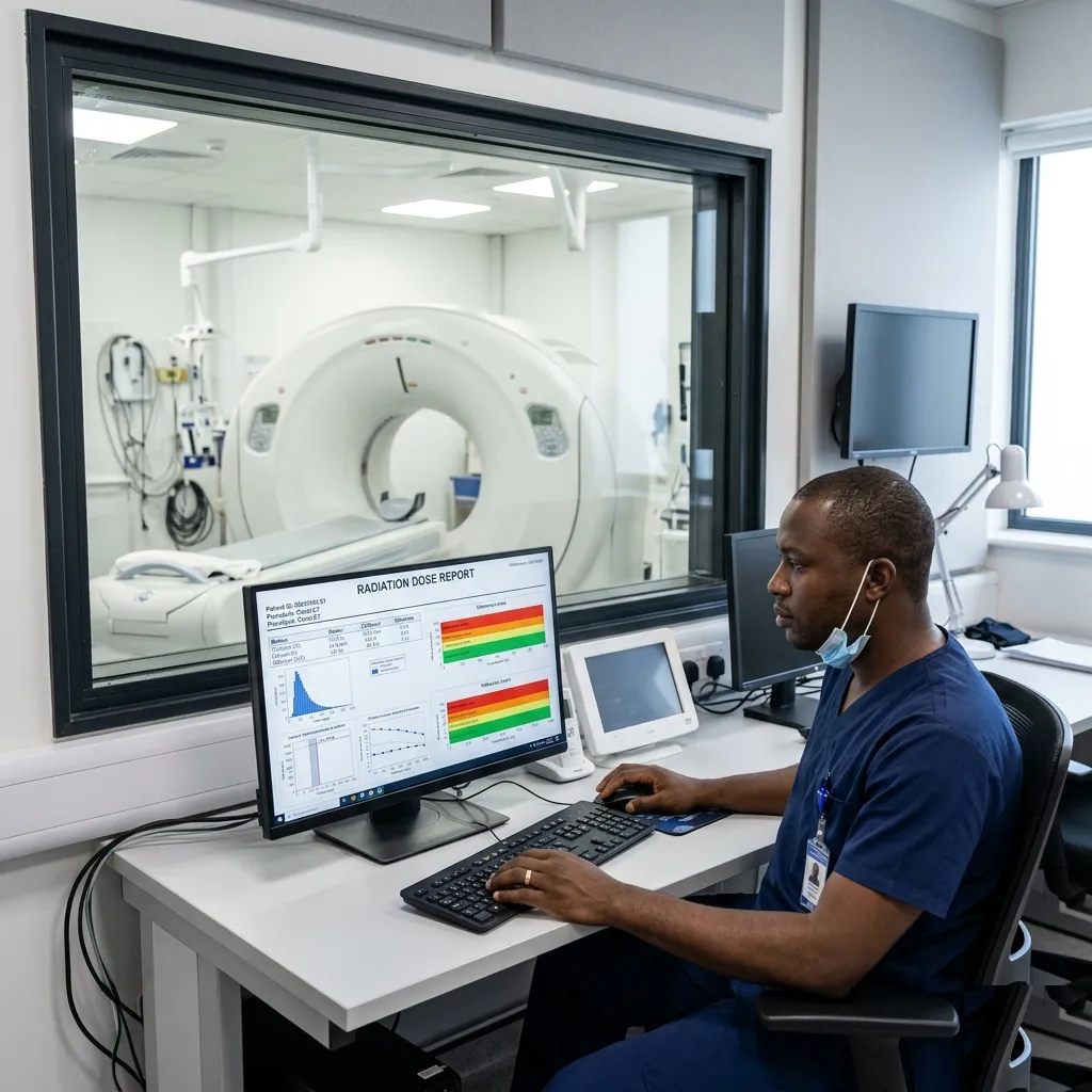
People who need repeated CT scans often worry that radiation is building up inside them. The radiation itself does not stay in the body, and a CT scan does not make you radioactive. What adds up is the exposure received across examinations.

CT dose is not one fixed number

A CT scan generally uses more radiation than a standard X-ray, but the dose varies widely. A brain CT, chest CT, and abdomen and pelvis CT do not give the same exposure. Dose also changes with the scan protocol, the patient's size, and whether more than one phase is required.

This is why a single percentage cannot accurately describe the cancer risk from every CT scan. Age, sex, body area, and total exposure all affect risk estimates, and there is uncertainty when very small risks are calculated.

Why doctors still request repeat CT

A repeat scan may answer a new question or show how something has changed. In cancer care, for example, it may show whether treatment is working. In an emergency, it may identify bleeding, a blood clot, or an injury that needs immediate treatment.

The possible long-term risk from radiation has to be weighed against the risk of missing or delaying an important diagnosis now.

How to avoid repetition that does not help

Keep your previous reports and images where possible, especially if you use more than one hospital or imaging centre. A report is useful, but the actual images may be needed for comparison.

You can also ask:

  • What question will this CT answer?
  • Can a recent scan be used instead?
  • Could ultrasound or MRI answer the same question?
  • Will the scan use a protocol adjusted for my age and size?

An alternative is only useful when it can answer the clinical question well enough. MRI and ultrasound do not use ionising radiation, but they cannot replace CT in every situation.

Children need extra attention

Children are more sensitive to radiation and have more years ahead in which a radiation-related effect could appear. CT protocols should be adjusted for a child's size, and the examination should be limited to what is needed.

The bottom line

There is no universal number of CT scans that becomes unsafe. Each examination should have a clear reason, use an appropriate protocol, and add information that can affect care.

Radiology education only

RadFAQS explains radiology terms, scan preparation, and what patients commonly experience. It is not a diagnosis, treatment plan, or replacement for your referring doctor, radiologist, or care team. RadFAQS does not monitor this site for emergencies and cannot respond in real time. If symptoms are severe, worsening, or urgent, do not wait for a reply here — contact a healthcare professional or emergency service immediately.

Related FAQ guides

mri

Breast MRI

A Breast MRI is a highly specialized imaging scan used to capture incredibly detailed pictures of breast tissue. It is frequently used alongside mammograms and ultrasounds to evaluate breast health.

mri

Fetal MRI

A Fetal MRI is a detailed scan of an unborn baby, usually performed in the second or third trimester to clarify findings seen on a prenatal ultrasound. It is safe, uses no radiation, and gives doctors crucial information for planning the rest of the pregnancy and delivery.

ultrasound

Musculoskeletal (MSK) Ultrasound

A Musculoskeletal (MSK) Ultrasound provides a real-time, moving picture of your muscles, tendons, ligaments, and joints. Unlike a static X-ray, this test allows your doctor to see how your internal structures move and stretch while you actively use them.

mri

TMJ MRI (Temporomandibular Joint)

A TMJ MRI focuses on the small joints in front of each ear that let you open, close, and move your jaw. It is the best scan for identifying jaw pain, clicking, locking, or disc problems that cannot be seen on a routine X-ray.

Related dictionary terms

Procedure

CT Scan

A CT scan uses X-rays and computer processing to create detailed cross-sectional images of the body.

Procedure

MRI

MRI uses magnets and radio waves to create detailed images, especially of the brain, spine, joints, and soft tissues.

Procedure

Ultrasound

Ultrasound uses sound waves to create real-time images of organs, soft tissues, pregnancy, and blood flow.

Procedure

X-ray

An X-ray is a quick imaging test that uses a small dose of radiation to show bones, parts of the chest, and some abdominal conditions.