Questions about this scan?
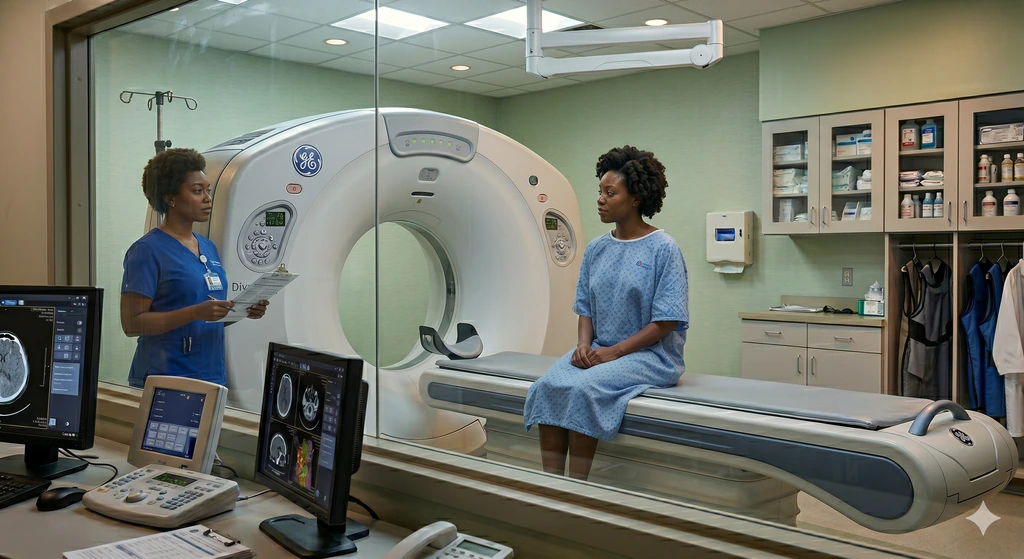
CT Scan
Computed Tomography (CT) uses X-rays combined with advanced computer processing to produce cross-sectional images of the body. CT scans can quickly and accurately show bones, soft tissues, blood vessels, and organs.
A CT Brain scan is one of the most commonly performed neuroimaging procedures in Radiology. It uses X-rays and computer processing to create highly detailed images of the brain and skull.
A CT Brain Angiography (CTA Brain) creates detailed maps of the blood vessels inside your head. It uses X-rays and computer processing to capture clear, 3D images of your cerebral circulation.
A CT Craniofacial scan captures clear, 3D-like images of the bones in your face and skull. It is a critical tool for mapping out the intricate bones of the cheeks, jaw, and eye sockets.
A CT IAM focuses tightly on the tiny, complex bony canals inside your skull. It provides a sharp look at the pathways that carry your hearing and balance nerves from your inner ear to your brain.
A CT Neck scan uses X-rays and special computer processing to produce detailed cross-sectional images of the neck region. It helps doctors visualize the soft tissues, airway, blood vessels, glands, lymph nodes, and cervical spine bones.
CT Paranasal Sinuses (often called CT PNS) is a specialized CT scan used to get detailed images of the paranasal sinuses. The paranasal sinuses are small, air-filled spaces in the bones around your nose that connect with the back of your throat.
A CT Temporomandibular joint (TMJ) scan focuses on the hinge joints connecting your jaw to your skull. It uses X-rays and special computer processing to produce clear, detailed pictures of the bones and their alignment.
A CT Chest scan is an imaging test that uses X-rays and computer processing to produce detailed cross-sectional images of the lungs, heart, airways, blood vessels, and chest wall.
A CT Spine scan uses X-rays and computers to produce crisp, cross-sectional pictures of your spine. It is an excellent tool for providing a pinpoint view of the bones, joints, and structures supporting your back and neck.
A CTPA (Computed Tomography Pulmonary Angiography) acts as a swift, targeted search for blockages in your lungs. It is a critical, life-saving test used to quickly map out the pulmonary arteries.
A High-Resolution CT (HRCT) Chest scan is like looking at a book page by page, rather than staring at the closed cover. It provides incredibly crisp, paper-thin slices of your lung tissue to uncover the root cause of chronic breathing issues.
A cardiac calcium scoring CT scan is a quick, non-contrast scan that measures the amount of calcified plaque in your heart's arteries. It helps assess your risk of developing a future heart attack.
A CT Cardiac scan is a non-invasive imaging test that uses X-rays and advanced computer processing to create detailed pictures of the heart.
A CT Coronary Angiography (CTCA) is a special type of CT scan that focuses on the coronary arteries, the blood vessels that supply the heart muscle. Using X-rays and contrast dye, CTCA produces detailed pictures that help doctors detect narrowing, blockages, and calcium buildup.
A CT Lower Limb Angiography maps out the blood vessels in your legs, providing a clear picture from your abdomen all the way down to your toes. It uses X-rays and computer processing to create detailed cross-sectional images of your vascular system.
A CT Upper Limb Angiography focuses on the blood vessels in your arms, from your shoulders down to your fingertips. It uses X-rays and computers to produce clear, 3D images of your arteries and veins.
A CTA Carotid (Computed Tomography Angiography of the Neck) is an imaging test that focuses on the major arteries in your neck that supply blood to your brain.
A CT Abdomen scan uses X-rays and computer processing to create detailed cross-sectional images of the abdominal organs including the liver, stomach, pancreas, kidneys, spleen, intestines, and blood vessels.
A CT Abdomen & Pelvis scan is a combined imaging study that examines your abdomen and pelvis in a single session. This allows doctors to see everything from the diaphragm to the pelvic floor, making it a very useful tool for diagnosing pain, infection, trauma, tumors, and other conditions that may involve multiple regions.
A CT Pancreas scan, often called a pancreatic protocol CT, is a targeted imaging test that focuses heavily on your pancreas and the surrounding digestive structures.
A CT Urogram is a special type of CT scan that focuses on the urinary tract. This includes the kidneys, ureters (the tubes that carry urine from the kidneys to the bladder), and the bladder itself.
Everything patients commonly ask about CT scans — what to expect, how long it takes, and whether it's safe.
A CT aortogram is a specialized scan that uses X-rays and contrast dye to create detailed images of your aorta. It helps detect dangerous conditions like aortic aneurysms or tears (dissections).
A CT Colonoscopy, also known as Virtual Colonoscopy, is a special type of CT scan used to examine the large intestine (colon and rectum). It uses X-rays and computer technology to create detailed 3D images of the inside of the bowel.
CT enterography uses oral contrast to distend the small intestine and CT imaging to assess its wall and surrounding tissues. It can help doctors look at an area of the digestive tract that is otherwise very hard to see.
A CT of the temporal bone is a high-resolution scan that examines the delicate structures of your middle and inner ear. It helps investigate hearing loss, chronic ear infections, or dizziness.
A CT Orbit scan is a special type of CT examination focused on the eye sockets (orbits) and the structures around them. It uses X-rays and computer processing to create detailed images of the bones, muscles, blood vessels, and soft tissues in and around your eyes.
CT pelvimetry measures the bony pelvis, but routine pelvimetry is not recommended for deciding mode of birth because it does not reliably predict whether vaginal birth will be successful.
A CT Triple Phase Study is a dynamic imaging test that looks at your organs at three precise moments in time. It uses X-rays and computer processing to capture images as blood flows through different stages.
A dental Cone Beam CT (CBCT) is a specialized X-ray scan that produces detailed three-dimensional images of your teeth, jawbone, nerves, and sinuses. It is commonly used to plan dental implants or orthodontic treatments.