Questions about this scan?
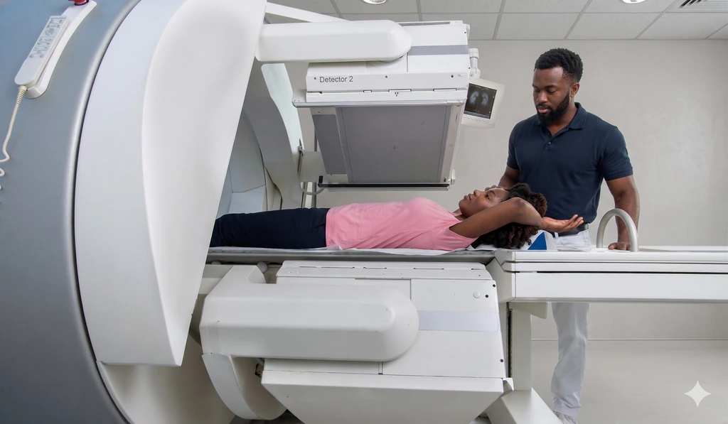
Nuclear Medicine
Nuclear medicine uses small amounts of carefully selected tracer and special cameras to show how organs and tissues are functioning. It is especially useful when doctors need answers about blood flow, drainage, organ performance, infection, inflammation, or cancer activity.
A bone scan is a nuclear medicine test that helps doctors look for changes in bone activity. It is often used for unexplained bone pain, infection, fractures, or to check whether cancer has spread to the bones.
A DMSA renal scan is a specialized nuclear medicine test used to examine the physical structure and scarring of your kidneys. It is commonly ordered for children with recurrent urinary tract infections.
A gastric emptying study is a nuclear medicine test that shows how quickly or slowly food moves through your stomach. It is often used when doctors suspect delayed or rapid stomach emptying.
A HIDA scan is a nuclear medicine test that helps doctors see how bile moves through the liver, gallbladder, bile ducts, and small intestine. It is often used to evaluate gallbladder function and some types of biliary blockage or leak.
A MAG3 renal scan is a specialized nuclear medicine test used to evaluate how well your kidneys drain urine. It helps detect blockages in the urinary tract.
A MUGA scan is a nuclear medicine heart test that measures how well your heart pumps blood. It is often used to assess ejection fraction, especially when doctors want to monitor heart function closely.
A myocardial perfusion scan is a nuclear medicine test that evaluates blood flow through your heart muscle at rest and during exercise. It helps detect blocked or narrowed coronary arteries.
Nuclear medicine uses tiny amounts of radioactive tracer and special cameras to show how organs and tissues are working. It helps doctors answer questions that ordinary X-rays, ultrasound, CT, or MRI may not answer on their own.