
RadFAQS
Scan GuidesDictionaryFind a CenterTalk to a Radiographer
Find/Book
HomeScan GuidesDictionaryFind a CenterTalk to a RadiographerBooking Follow-Up
RadFAQS

Nigeria's radiology directory - helping patients find listed diagnostic centers, understand their scans, and take control of their health.

Modalities

  • CT Scan
  • MRI
  • DEXA
  • X-ray
  • Ultrasound
  • Fluoroscopy
  • Mammography
  • Nuclear Medicine
  • Interventional Radiology
  • Radiotherapy

Platform

  • Find/Book a Scan
  • Talk to a Radiographer
  • Booking Follow-Up
  • Volunteer

Portals

  • Center Login

Stay in the Know

Get updates on new centers, health tips, and platform news.

support@RadFAQS.comWhatsApp Us
© 2026 RadFAQS. All rights reserved.
Privacy PolicyTerms of UseRefund PolicyAbout
Back to Articles
Radiology Terms & Requests

What 'Prior Images for Comparison' Means

SM
Written by Sangodoyin Maryam, B.Sc Radiography
·
Medically reviewed by Olusegun Samuel Faith, M.Sc (Medical Imaging), MPH, PgDip (MRI)· Last reviewed 27 Mar 2026
What 'Prior Images for Comparison' Means

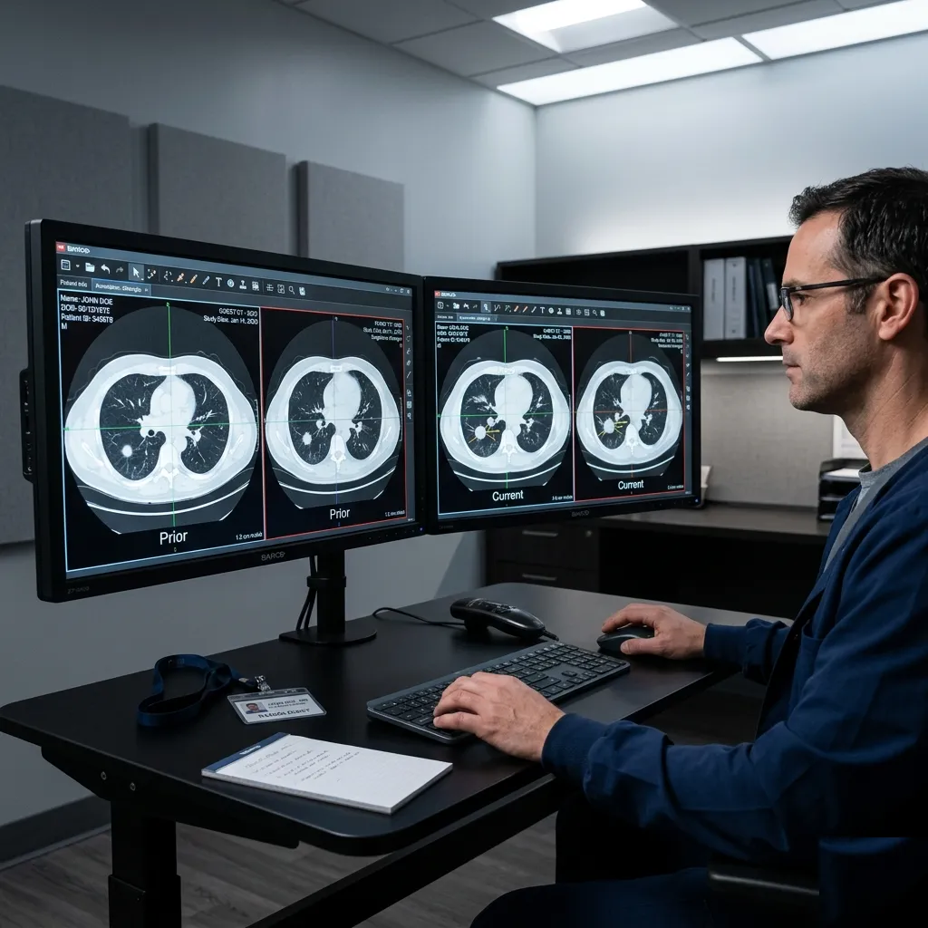
You may hear a center ask for prior images for comparison or see earlier studies mentioned in a report.

That is not administrative fussiness. It is often one of the most useful parts of the interpretation.

Why comparison matters so much

Radiology is not always about what a finding is in isolation. It is often about change over time.

Comparing with older scans helps answer questions like:

  • Is this new?
  • Has it grown?
  • Has it stayed stable for years?
  • Is treatment working?

The exact same image finding can feel much more reassuring or much more concerning depending on that timeline.

When prior studies are especially valuable

Comparison is particularly helpful for:

  • Lung nodules
  • Breast findings
  • Chronic spine changes
  • Cancer follow-up
  • Cysts or masses that have been watched before

Why the old report alone is not always enough

Sometimes the written report helps. But sometimes the actual images are even more valuable.

Different radiologists may describe the same thing slightly differently. Seeing the old images directly allows a more confident side-by-side judgment.

What patients can do

If you have previous scans from another center, bring:

  • The report
  • The image disc or digital link, if available

That small effort can save repeat imaging and reduce uncertainty.

Worth remembering

Stable over time is one of the most reassuring phrases in radiology, and it is only possible when something is available for comparison.

The bottom line

Prior images are not just background paperwork. They help the radiologist understand whether a finding is standing still, evolving, or resolving, which often changes what happens next.

Radiology education only

RadFAQS explains radiology terms, scan preparation, and what patients commonly experience. It is not a diagnosis, treatment plan, or replacement for your referring doctor, radiologist, or care team. RadFAQS does not monitor this site for emergencies and cannot respond in real time. If symptoms are severe, worsening, or urgent, do not wait for a reply here — contact a healthcare professional or emergency service immediately.

Related FAQ guides

interventional-radiology

Soft Tissue Mass Biopsy

A Soft Tissue Mass Biopsy evaluates unexplained lumps under the skin, in the fat, or deep inside a muscle using ultrasound or CT guidance to precisely identify what the mass is made of.

mri

Soft Tissue Mass MRI

A Soft Tissue Mass MRI is a detailed scan of a lump or growth in the muscle, fat, or other soft tissues. It is used to characterise the lump, distinguish benign growths from sarcomas, and guide biopsy or surgery.

fluoroscopy

Cystography

A cystogram is a special X-ray study of the bladder using contrast dye. It is used to detect bladder leaks, fistulas, abnormal pouches, and reflux from the bladder back up toward the kidneys.

mri

Adrenal MRI

An Adrenal MRI is a focused scan of the small glands that sit above each kidney. It is most often used to characterise a mass found unexpectedly on another scan, and to tell harmless adenomas apart from lesions that need further attention.

Related dictionary terms

Term

Cyst

A cyst is a fluid-filled sac that can occur in many parts of the body.

Term

Mass

Mass is a broad imaging term for a lump or abnormal growth seen in an organ or tissue.

Term

Nodule

A nodule is a small rounded or irregular area seen in tissue, commonly in the lungs, thyroid, liver, or elsewhere.