Cervical spondylosis refers to age-related wear-and-tear changes in the neck portion of the spine. It is common, especially as people get older, and many imaging reports mention it.
That does not automatically mean severe disease. The real question is whether the changes are mild background aging or whether they are compressing nerves or the spinal cord.
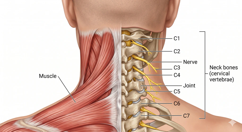
What radiology helps show
Imaging may be used to assess:
- Disc space narrowing
- Bone spurs
- Alignment changes
- Narrowing where nerves exit
- Spinal canal narrowing
- Possible cord compression
Doctors may use:
- X-rays for basic bony alignment and degenerative change
- MRI when nerve symptoms, weakness, numbness, or possible cord involvement are concerns
Why symptoms matter so much
Some people have obvious spondylotic changes on imaging and very little pain. Others have smaller-looking changes but more symptoms.
That is why management is not based on the scan alone.
How it is often managed
Depending on severity, care may include:
- Pain relief strategies
- Physiotherapy
- Activity modification
- Posture and ergonomic changes
- Specialist evaluation if nerve or cord symptoms appear significant
- Surgery in selected cases where compression is serious
When imaging findings become more important
Radiology takes on extra significance if there are:
- Arm weakness
- Numbness
- Hand clumsiness
- Balance trouble
- Signs suggesting spinal cord involvement
Those are the situations where MRI is often especially useful.
Good to remember
Cervical spondylosis is common on scans. What matters most is how the imaging findings line up with symptoms and neurological exam findings.
The bottom line
Radiology helps show the structural side of cervical spondylosis, but the management plan is built from both the scan and the patient’s actual symptoms, function, and exam.