Osteoarthritis is one of the most common joint conditions people encounter. It is a wear-related process affecting cartilage, bone, and the overall joint structure.
People often think of it simply as "arthritis," but that word covers several different conditions. Osteoarthritis is the common degenerative type.
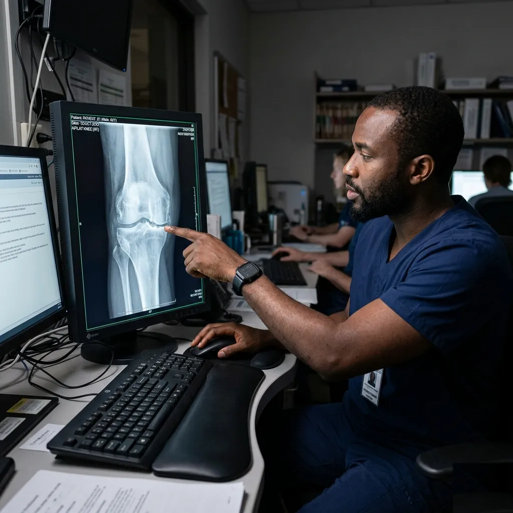
What imaging can show
Radiology may reveal features such as:
- Joint space narrowing
- Bone spurs
- Subchondral sclerosis
- Cysts and chronic wear-related changes
X-rays are often the first imaging test used. In some situations, MRI may be requested if there is a need to assess soft tissue, cartilage, or to rule out another issue.
Why the scan is not the whole story
Some patients have obvious osteoarthritic changes on imaging and only modest symptoms. Others have substantial pain with less dramatic imaging.
That is why doctors do not treat the picture alone. They treat the person.
How osteoarthritis is commonly managed
Management often includes:
- Exercise and physiotherapy
- Weight management where relevant
- Pain relief strategies
- Joint injections in selected cases
- Activity modification
- Surgery, including joint replacement, in more advanced disease
The exact plan depends on the affected joint, symptom severity, mobility, and how much daily life is being disrupted.
When imaging becomes especially helpful
Radiology is particularly useful when the team needs to:
- Confirm the pattern fits osteoarthritis
- Assess severity
- Rule out fracture or another joint problem
- Plan intervention or surgery
Helpful perspective
Imaging can confirm osteoarthritis, but management decisions are usually guided by both the scan and how much pain, stiffness, and function loss the patient is actually experiencing.
The bottom line
Osteoarthritis is common, and imaging helps show the structural side of it clearly. But the role of radiology is to support good management decisions, not to replace the broader clinical picture.