
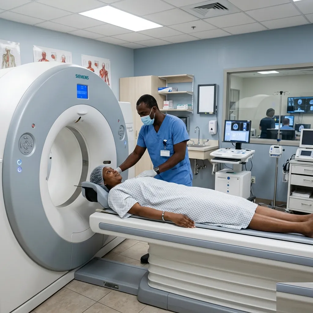
A PET-CT may be requested after a cancer diagnosis, during treatment, or when a doctor needs to check for recurrence. It combines two kinds of information in one examination.
The CT part shows structure: the size and location of organs, lymph nodes, and other tissues. The PET part shows how a tracer is being taken up in the body. The most commonly used tracer, FDG, behaves in a similar way to glucose.
What the bright areas mean
Areas that take up more tracer may appear brighter on the PET images. Some cancers have increased uptake because their cells are more metabolically active.
Bright does not automatically mean cancer. Infection, inflammation, healing tissue, normal organs, and muscle activity can also take up FDG. Some cancers take up very little. This is why the radiologist interprets the pattern alongside the CT images, your medical history, and earlier scans.
Where PET-CT may help
Depending on the cancer type, PET-CT may help doctors:
- see whether disease has spread;
- choose an area for biopsy;
- plan radiotherapy or other treatment;
- assess how a cancer has responded to treatment; or
- investigate whether cancer may have returned.
PET-CT is not needed for every cancer. It also does not decide treatment on its own. Biopsy results, blood tests, symptoms, and other scans may be just as important.
Why timing matters
Recent surgery, radiotherapy, chemotherapy, infection, or high blood sugar can affect tracer uptake. The oncology and imaging teams choose the timing of the scan carefully so the result is easier to interpret.
The bottom line
PET-CT helps doctors connect activity with location. Its value depends on the type of cancer and the question the care team is trying to answer, not simply on it being a more advanced scan.