
RadFAQS
Scan GuidesDictionaryFind a CenterTalk to a Radiographer
Find/Book
HomeScan GuidesDictionaryFind a CenterTalk to a RadiographerBooking Follow-Up
RadFAQS

Nigeria's radiology directory - helping patients find listed diagnostic centers, understand their scans, and take control of their health.

Modalities

  • CT Scan
  • MRI
  • DEXA
  • X-ray
  • Ultrasound
  • Fluoroscopy
  • Mammography
  • Nuclear Medicine
  • Interventional Radiology
  • Radiotherapy

Platform

  • Find/Book a Scan
  • Talk to a Radiographer
  • Booking Follow-Up
  • Volunteer

Portals

  • Center Login

Stay in the Know

Get updates on new centers, health tips, and platform news.

support@RadFAQS.comWhatsApp Us
© 2026 RadFAQS. All rights reserved.
Privacy PolicyTerms of UseRefund PolicyAbout
Back to Articles
Understanding Modalities

What a Mammogram Actually Checks

TO
Written by Taiwo Oluwayemisi, B.Sc Radiography
·
Medically reviewed by Olusegun Samuel Faith, M.Sc (Medical Imaging), MPH, PgDip (MRI)· Last reviewed 6 Apr 2026
What a Mammogram Actually Checks

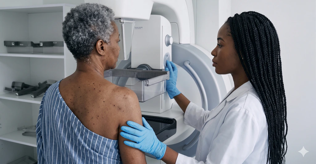
A mammogram is a specialized X-ray of the breast. It is designed to find changes that may be too small to feel by hand.

That matters because breast imaging is often about catching subtle findings early, when the next step can be simpler and outcomes can be better.

What it can show

A mammogram may help detect:

  • Small masses
  • Calcifications
  • Architectural distortion
  • Asymmetry between breasts

Some of these findings are harmless. Some need follow-up. The scan itself is about spotting patterns that deserve a closer look.

Screening versus diagnostic mammography

Not every mammogram is ordered for the same reason.

  • A screening mammogram is for routine early detection when there is no specific new complaint.
  • A diagnostic mammogram is more targeted and is usually done when there is a symptom or an abnormal earlier study.

That difference affects the images taken and sometimes how quickly you get feedback.

Why compression is used

Compression is uncomfortable for some patients, but it serves a purpose. It spreads the breast tissue out, reduces blur, and helps the machine capture more useful detail with less radiation.

It is not there to punish you. It is there to improve the exam.

When extra imaging may still be needed

A mammogram does not always end the story.

Sometimes the radiologist may recommend:

  • Extra mammographic views
  • Breast ultrasound
  • Breast MRI

That does not automatically mean cancer. It often means the first set of images raised a question that needs a better answer.

Important context

Dense breast tissue can make mammograms harder to interpret. That is one reason some people need additional imaging even when nothing dangerous is ultimately found.

The bottom line

A mammogram is a focused tool for detecting breast changes early and guiding what should happen next. It is one of the most important imaging tests in preventive care, even when the process itself is not anyone's favorite day.

Radiology education only

RadFAQS explains radiology terms, scan preparation, and what patients commonly experience. It is not a diagnosis, treatment plan, or replacement for your referring doctor, radiologist, or care team. RadFAQS does not monitor this site for emergencies and cannot respond in real time. If symptoms are severe, worsening, or urgent, do not wait for a reply here — contact a healthcare professional or emergency service immediately.

Related FAQ guides

mammography

Mammography

Mammography is a branch of Radiology that involves the use of low-energy X-rays to see the inside of the breasts. This investigation is of growing popularity because it can be used to routinely check the breast for growths, lesions or calcifications without much associated risks.

mri

Soft Tissue Mass MRI

A Soft Tissue Mass MRI is a detailed scan of a lump or growth in the muscle, fat, or other soft tissues. It is used to characterise the lump, distinguish benign growths from sarcomas, and guide biopsy or surgery.

mri

Breast MRI

A Breast MRI is a highly specialized imaging scan used to capture incredibly detailed pictures of breast tissue. It is frequently used alongside mammograms and ultrasounds to evaluate breast health.

ultrasound

Breast Ultrasound

A Breast Ultrasound uses sound waves to evaluate breast tissue, often acting as a companion test to a standard mammogram. It helps doctors take a much closer look at a specific lump or area of concern to figure out exactly what it is.

Related dictionary terms

Term

Calcification

Calcification means calcium has collected in tissue and become visible on imaging.

Procedure

Mammography

Mammography is a specialized X-ray used for breast screening and diagnostic assessment.

Term

Mass

Mass is a broad imaging term for a lump or abnormal growth seen in an organ or tissue.

Procedure

MRI

MRI uses magnets and radio waves to create detailed images, especially of the brain, spine, joints, and soft tissues.