
RadFAQS
Scan GuidesFind a CenterTalk to a Radiographer
Find/Book
HomeScan GuidesFind a CenterTalk to a RadiographerBooking Follow-Up
RadFAQS

Nigeria's radiology directory - helping patients find listed diagnostic centers, understand their scans, and take control of their health.

Modalities

  • CT Scan
  • MRI
  • DEXA
  • X-ray
  • Ultrasound
  • Fluoroscopy
  • Mammography
  • Nuclear Medicine
  • Interventional Radiology
  • Radiotherapy

Platform

  • Find/Book a Scan
  • Talk to a Radiographer
  • Booking Follow-Up
  • Dictionary
  • Volunteer

Portals

  • Center Login

Stay in the Know

Get updates on new centers, health tips, and platform news.

support@RadFAQS.comWhatsApp Us
© 2026 RadFAQS. All rights reserved.
Privacy PolicyTerms of UseRefund PolicyAbout
Back to Articles
Modality Basics

X-Ray Safety and Radiation: What You Need to Know

OA
Written by Oluwatobiloba Akinnusi, B.Sc Radiography
·
Medically reviewed by Olusegun Samuel Faith, M.Sc (Medical Imaging), MPH, PgDip (MRI)· Last reviewed 10 Jun 2026
X-Ray Safety and Radiation: What You Need to Know

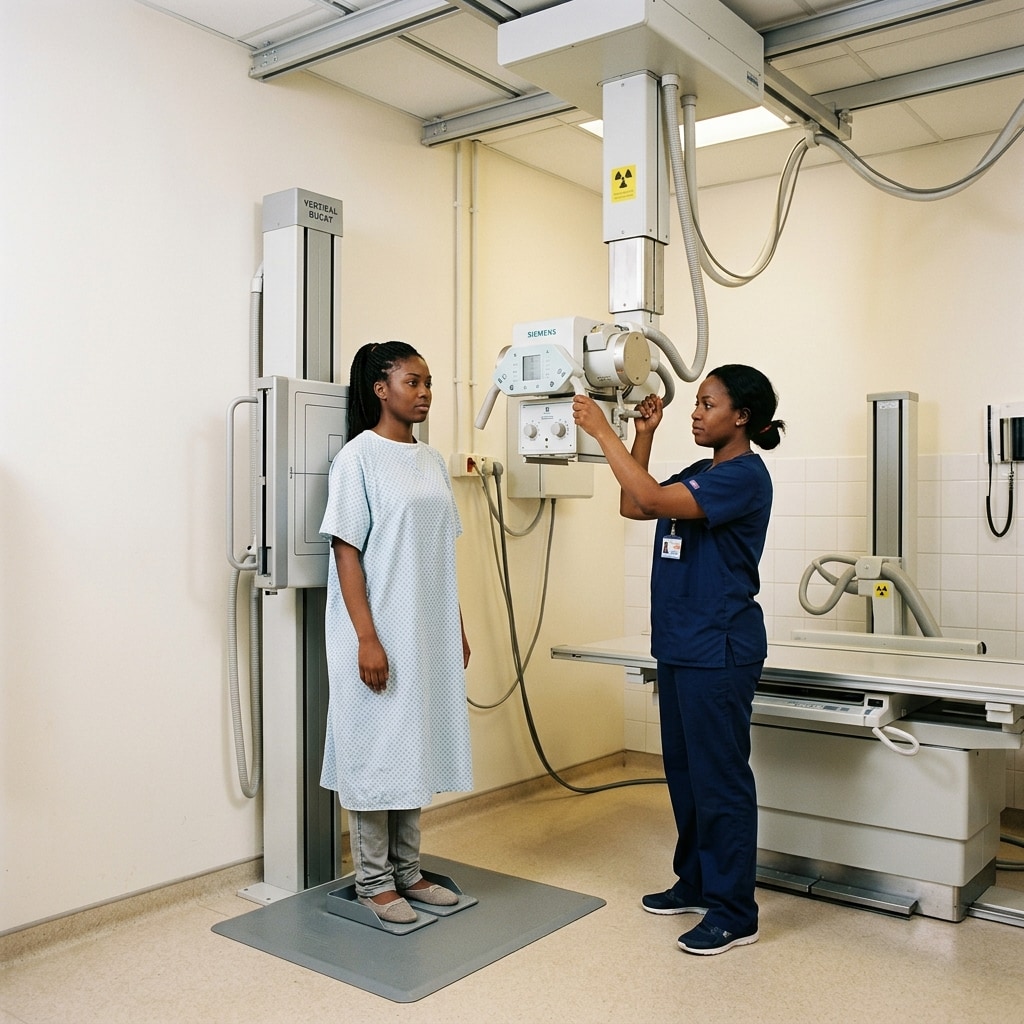
Standard X-rays are the foundation of medical imaging. Whether checking for a broken bone in your foot or screening for chest infections, X-rays are fast and effective. However, because they use ionizing radiation, many patients feel anxious about their safety.

The level of radiation is very low

The amount of radiation used in a standard diagnostic X-ray is extremely small. To make this relatable, scientists compare medical radiation to the natural background radiation we receive every day from the earth and outer space.

  • A standard chest X-ray exposes you to about 0.1 mSv of radiation. This is equivalent to about 10 days of natural background radiation.
  • An X-ray of a limb (like a hand or foot) uses even less—less than a single day of natural background radiation.

Because the dose is so small, the risk of developing any health issues from a standard diagnostic X-ray is practically zero.

How the team keeps you safe

Radiographers follow a safety principle called ALARA, which stands for "As Low As Reasonably Achievable." This means they use the absolute minimum amount of radiation needed to get a clear, diagnostic picture.

  • Lead shielding: The radiographer may place a lead apron or shield over your abdomen or pelvis to protect your reproductive organs from scattered radiation.
  • Collimation: The X-ray machine is adjusted so the beam only hits the specific area being studied (for example, just your ankle), keeping the rest of your body completely safe.
  • Behind the screen: The radiographer stands behind a lead-shielded wall or screen during the exposure. They do this because they perform dozens of scans every day, and cumulative exposure must be prevented for staff.

Preparing for your X-ray

X-rays require almost no preparation. You can eat and drink normally. The most important step is to wear loose, comfortable clothing and remove any metal objects from the area being scanned. Metals block X-rays and show up as white shapes, which can hide fractures or infections on the image.

Pregnancy guidelines

If you are pregnant or think you might be, always inform the radiographer before the scan. While standard X-rays of the limbs or chest expose the womb to almost zero radiation, the team will take extra precautions (like double shielding) or discuss using ultrasound instead.

A comforting close

Standard X-rays are exceptionally safe and provide your referring doctor with vital information in minutes. The risk of missing a fracture or infection far outweighs the negligible radiation risk of a diagnostic X-ray.

Radiology education only

RadFAQS explains radiology terms, scan preparation, and what patients commonly experience. It is not a diagnosis, treatment plan, or replacement for your referring doctor, radiologist, or care team. RadFAQS does not monitor this site for emergencies and cannot respond in real time. If symptoms are severe, worsening, or urgent, do not wait for a reply here — contact a healthcare professional or emergency service immediately.

Related FAQ guides

xray

Chest X-Ray

A chest X-ray is a quick scan that uses a small amount of radiation to take pictures of your lungs, heart, ribs, and chest bones. Doctors commonly request it for cough, chest pain, breathing difficulty, infections, and chest injuries.

xray

Clavicle (Collarbone) X-Ray

A clavicle X-ray is a quick scan that uses a small amount of radiation to show the collarbone. Doctors commonly request it after a fall, shoulder injury, visible swelling, pain, or suspected collarbone fracture.

xray

Elbow X-Ray

An elbow X-ray is a quick scan that uses a small amount of radiation to show the bones around the elbow joint. It is commonly requested after falls, swelling, pain, stiffness, suspected fracture, or reduced arm movement.

xray

Foot X-Ray

A foot X-ray is a quick scan that uses a small amount of radiation to show the bones of the foot. It is often requested after injury, pain, swelling, difficulty walking, suspected fracture, or follow-up after treatment.

Related dictionary terms

Procedure

X-ray

An X-ray is a quick imaging test that uses a small dose of radiation to show bones, parts of the chest, and some abdominal conditions.

Pathology

Fracture

A fracture is a break in a bone, ranging from a fine crack to a complete break with displacement.

Term

Radiograph

A radiograph is the formal name for an X-ray image.

Procedure

Ultrasound

Ultrasound uses sound waves to create real-time images of organs, soft tissues, pregnancy, and blood flow.